Established, 2004 in the UK
CPD accredited, online courses for doctors in all of the major specialties

Body Imaging: Managing the Incidental Findings

Wednesday 15 and Thursday 16 and Friday 17 May 2024

Incidental Findings course banner
Edit Content

DAY ONE: THORACIC IMAGING

Wednesday 15 May 2024

All times are GMT+1

09:50 – 10.00

Introduction to Webex and PACS

Jack Hodson

Online and IT Services Manager, Infomed Online.

10.00 – 11.20

Musculoskeletal

Picture of Dr David Elias

Dr David Elias

Consultant MSK Radiologist, King’s College Hospital

11.20 - 11.40

Break

11.40 – 12.40

Heart and Vascular Structures

Dr David P. Munger

Assistant Professor of Radiology, Dartmouth-Hitchcock Medical Center, USA

12.40 – 13.30

Lunch

13.30 – 14.30

Thyroid

Picture of Dr Ichiro Ikuta

Dr Ichiro Ikuta

Mayo Clinic Arizona, Senior Associate Consultant , Department of Radiology, Section of Neuroradiology

14.30 – 15.30

Lungs

Picture of Dr Matthew Maeder

Dr Matthew Maeder

Assistant Professor of Radiology, Dartmouth-Hitchcock Medical Center, USA

15.30 – 16.00

Lunch

16.00 - 17.00

Mediastinum

Dr Cara Owens

Consultant Radiologist, The Royal Marsden Hospital, London, UK

DAY TWO: UPPER ABDOMEN

Thursday 16 May 2024

All times are GMT+1

09.30 – 10.10

Spleen

Picture of Dr Christopher Harvey

Dr Christopher Harvey

Consultant radiologist, Imperial NHS Trust, London

10.10 – 10.50

Lymph nodes

Picture of Dr Ken Courtney

Dr Ken Courtney

Consultant Interventional Radiologist, King’s College Hospital

10.50 – 11.10

Break

11.10 – 12.20

Adrenal Glands

Picture of Dr Anju Sahdev

Dr Anju Sahdev

Radiology Consultant and Director for Research at St Bartholomew's Hospital

12.20 – 13.30

Gallbladder and Bile Ducts

Picture of Dr Priti Dutta

Dr Priti Dutta

Consultant Radiologist at the Royal Free Hospital, London, UK

13.30 – 14.20

Lunch

14.20 – 15.40

Liver

Picture of Dr Kevin Kalisz

Dr Kevin Kalisz

Assistant Professor at Duke University School of Medicine, USA

15.40 – 16.00

Break

16.00 – 17.00

Pancreas

Picture of Dr Mahan Mathur

Dr Mahan Mathur

Associate Professor of Radiology and Biomedical Imaging, Division Chief: Body Imaging, Yale School of Medicine, USA

DAY THREE: GI/GU

Friday 17 May 2024

All times are GMT+1

09.30 – 10.55

Gynaecology

Picture of Dr Nishat Bharwani

Dr Nishat Bharwani

Consultant Radiologist at Imperial College Healthcare NHS Trust and Honorary Clinical Senior Lecturer at Imperial College London, UK

10.55 – 11.15

Break

11.15 – 12.15

Urinary bladder

Dr Anish Raithatha

Consultant Radiologist, Australia

12.15 – 13.00

Kidneys/Ureters - Incidental Solid masses?

Dr Tim Yusuf

Consultant Radiologist, King's College Hospital London

13.00 – 13.45

Lunch

13.45 – 14.45

Kidneys

Dr Michele Bertolotto

Associate Professor of Radiology, Head of the Unit of Multiparametric Ultrasound, University of Trieste, Italy

14.45 – 15.15

Break

15.15 – 16.45

Incidental Findings in the GI tract

Picture of Dr Matthew Maeder

Dr Matthew Maeder

Assistant Professor of Radiology, Dartmouth-Hitchcock Medical Center, USA

Edit Content

The aim

By the end of the webinar, participants will have:

Edit Content

An international Faculty, listing experts with years of experience in reporting (and in disseminating skills and practical knowledge to peers) from leading British and American hospitals, namely:

Course director

Picture of Dr Matthew Maeder

Dr Matthew Maeder

Assistant Professor of Radiology, Divisions of Neuroradiology
and Abdominal Imaging, Dartmouth-Hitchcock Medical Center, New Hampshire

Dr. Matthew Maeder is an Assistant Professor of Radiology at the Dartmouth-Hitchcock Medical Center. He completed medical school at the University of Cincinnati, followed by Radiology residency training at the Lenox Hill Hospital in New York City. He is dual fellowship-trained in both Body MRI and Neuroradiology, both at Dartmouth. His interests include diseases of the central nervous system, head and neck, as well as the liver and prostate. Dr. Maeder is experienced in a variety of procedures including CT-guided biopsies of the lungs, liver, and kidneys as well as spinal procedures including vertebroplasty, sacroplasty, facet joint synovial cyst rupture, and spine biopsy. He is currently on the faculty at Dartmouth specializing in Abdominal Imaging and Neuroradiology. He is the Director of the Body MRI fellowship.

Faculty members

Picture of Professor Paul Sidhu

Professor Paul Sidhu

Consultant Radiologist, Professor of Imaging Sciences and Clinical Director of Radiology, King's College Hospital London

Paul Sidhu is Professor of Imaging Sciences at King's College London and a Consultant Radiologist in the Department of Radiology at King' College Hospital.  He is Clinical Director of Radiology at King’s College Hospital, London.

He qualified from St. Mary’s Hospital Medical School (now part of Imperial College) in 1982, with Honours. He undertook his Radiology training at the Hammersmith Hospital and King's College Hospital and was appointed a Consultant Radiologist at King's College Hospital in 1996, with a role in Ultrasound and Interventional Radiology. He was made Professor of Imaging Sciences in 2012.

He has published extensively on many aspects of Ultrasound particularly in relation to male health and liver transplantation and has pioneered the introduction of contrast-enhanced ultrasound in the United Kingdom. He is recognised as an authority in the application of contrast-enhanced ultrasound in clinical practice.

He is the Editor of 8 books, and has published over 600 scientific articles, book chapters and conference abstracts. He lectures widely with over 500 presentations at national and international meetings.

He is currently the Editor of the European Journal of Ultrasound and previously the Deputy Editor of the British Journal of Radiology. He is an Associate Editor of Radiology.

He is past-President of the British Medical Ultrasound Society and the Past-President of the Section of Radiology of the Royal Society of Medicine. He is recent Past President of the European Federation of Societies in Ultrasound in Medicine and Biology.

His current research interests include the application of contrast enhanced ultrasound to the testis, radiation dose reduction in children and non-invasive ultrasound in the assessment of liver disease.

Picture of Dr Miriam Salib

Dr Miriam Salib

Dr Miriam Salib is a Consultant Radiologist at Barts Health NHS Trust, London, UK. She earned her medical degree from The University of Nottingham and undertook her radiology training at Imperial. Her specialist interest is in GU imagaing.

Picture of Dr Ichiro Ikuta

Dr Ichiro Ikuta

Senior Associate Consultant, Mayo Clinic Arizona

Picture of Dr Cara Owens

Dr Cara Owens

Dr Cara Owens is aconsultant radiologist at the Royal Marsden Hospital, London with special interests in thoracic malignancy and lymphoma. Following core radiology training at Chelsea and Westminster Hospital, London she specialised in chest imaging through the Thoracic Imaging Fellowship at the Royal Brompton Hospital, London. Cara has a strong research portfolio having obtained a PhD in molecular pathology from the University of Cambridge and she is currently completing my MDRes in imaging of myositis-associated fibrotic interstitial lung disease at Royal Brompton Hospital/Imperial College London. Cara relishes a challenge, whether it be work-related, learning a new language or trying to keep up with her daughters in the pool!

Picture of Dr. David P. Munger

Dr. David P. Munger

Assistant Professor of Radiology, Divisions of Interventional Radiology and Abdominal Imaging, Dartmouth-Hitchcock Medical Center, New Hampshire
He earned an undergraduate degree in Chemical Engineering with High Honors from the Worcester Polytechnic Institute. He then went on to medical school at the University of New England where he earned a concurrent Masters in Public Health. This was followed by radiology training at the Norwalk Hospital / Yale School of Medicine, where he completed subspecialty training in cardiovascular imaging. He is fellowship-trained in Interventional Radiology at the Dartmouth-Hitchcock Medical Center. His interests include diseases of the hepatobiliary system and genitourinary system, in addition to ongoing cardiothoracic diseases.

Picture of Dr Kevin Kalisz

Dr Kevin Kalisz

Assistant Professor of Radiology, Division of Abdominal Imaging, Duke University Medical Center, Durham, North Carolina 

Dr. Kalisz earned his Bachelor of Science in biomedical engineering and economics from Northwestern University and obtained a medical degree from Northwestern University Feinberg School of Medicine in Chicago, Illinois. He completed his residency training at University Hospital Cleveland Medical in Cleveland, Ohio where he also served as chief resident. Following residency, he completed fellowship training in abdominal imaging at Duke University Medical Center as well as cardiovascular imaging at Northwestern Memorial Hospital. He subsequently joined the Department of Radiology at Duke University Medical Center in the Abdominal Imaging Division. His clinical and academic interests include oncologic imaging, particularly rectal cancer imaging, dual energy CT, and cardiovascular CT and MRI.

Picture of Dr Mahan Mathur

Dr Mahan Mathur

Associate Professor of Radiology and Biomedical Imaging, Division Chief: Body Imaging,
Yale School of Medicine, USA

After receiving his medical degree from McGill University School of Medicine, Dr. Mahan Mathur completed his Diagnostic Radiology Residency from Yale-New Haven Hospital where he served as Chief Resident, followed by a Fellowship in Abdominal Imaging and Intervention at Massachusetts General Hospital.

Since 2013, he has served as faculty at the Yale School of Medicine, where he is an Associate Professor of Radiology and Biomedical Imaging and Chief of Body Imaging. In addition, he serves as an Associate Program Director for the Diagnostic Radiology Residency. Dr. Mathur is passionate about Radiology education and mentorship and has been the recipient of the RSNA honored educator award in 2017 as well as number departmental “Teacher” and “Mentor” of the year awards. While widely published across all aspects of Body Imaging, his scholarly output has focused on male genitourinary (GU) conditions and intravenous contrast agent guidelines.

Picture of Dr Anju Sahdev

Dr Anju Sahdev

Radiology Consultant and Director for Research at St Bartholomew's Hospital, London, UK

Dr Sahdev is a Uro-Gynae Radiology Consultant, a Reader in Diagnostic Imaging and Director for Imaging Research at St Bartholomew’s Hospital, Barts Health, London.

Having graduated from University of London in 1991, Dr Sahdev completed her Radiology training at University College Hospital and St Bartholomew’s Hospital, becoming a fellow of the Royal College of Radiologists in 1998.

Her radiological specialties are imaging in abdominal and pelvic oncology, particularly gynaecological, endocrine and uro-oncology diseases. 

Dr Sahdev practices at a high standard in ultrasound, CT, MRI and PET CT imaging.

She has authored and co-authored more than 85 peer reviewed publications, has authored 20 chapters in clinical and imaging textbooks and lectures regularly at national and international meetings.

Dr Sahdev is an associate editor for the British Journal of Radiology and Cancer Imaging and a reviewer for several peer review journals including Clinical Radiology, European Radiology and a member of the educational subcommittee of  the International Cancer Imaging Society. 

She is the gynaecological imaging module lead for the UK Radiology Integrated Training Initiative (R-ITI) e-learning programme.

Picture of Dr Nishat Bharwani

Dr Nishat Bharwani

Dr Nishat Bharwani is a Consultant Radiologist at Imperial College Healthcare NHS Trust and Honorary Clinical Senior Lecturer at Imperial College London. She qualified with honours from Guy’s, King’s & St Thomas’ Medical School in 2000 and undertook basic medical training in London and Brighton, obtaining membership of the Royal College of Physicians in 2003. She completed general Radiology training at St George's Hospital (2003-2008) and a fellowship in body MRI at Barts and The London NHS Trust (2008-2010), becoming a Fellow of the Royal College of Radiologists in 2008. Her radiological interests include gynaecological imaging, oncological imaging and urological imaging and she has published in these areas. Nishat is head of specialty for Gynaecological and Urological Imaging at Imperial College Healthcare NHS Trust.
Nishat is heavily involved in teaching as joint Training Programme Director for Radiology trainees at Imperial, joint academic training lead for the London School of Radiology, and regularly delivers lectures and workshops both nationally and internationally. She is an examiner for the Royal College of Radiologists FRCR 2a examination.

Picture of Dr Christopher Harvey

Dr Christopher Harvey

Consultant Radiologist, Imperial Healthcare NHS Trust, London

Dr. Harvey qualified from Charing Cross and Westminster Hospital Medical School having obtained an intercalated BSc in Physiology. He initially trained in medicine and obtained MRCP.

Dr. Harvey then trained in radiology at The Middlesex/UCL and Hammersmith Hospitals before becoming MRC research fellow at Hammersmith Hospital.

He became a consultant at Hammersmith Hospital in 2001 as well as head of ultrasound. Dr Harvey has published over 50 peer reviewed papers, three books, several chapters, as well as written or co-authored many articles on a wide spectrum of radiological subjects.

He is actively involved in teaching on several national FRCR courses. Dr. Harvey holds national as well as international editorial positions and is responsible for refereeing papers and organising courses/seminars.

Dr. Harvey’s clinical interests are general cross sectional imaging: CT colonography, CT coronary angiography, transrectal ultrasound and prostatic biopsies, carotid Doppler ultrasound as well as the renal/urogenital tract and hepatobiliary system.

His research interests include ultrasound, ultrasound contrast agents and functional imaging.

His special interests include General abdominal imaging, CT colonoscopy, CT coronary angiography, ultrasound carotids, transrectal ultrasound and prostate biopsies, ultrasound contrast agents

Picture of Dr Priti Dutta

Dr Priti Dutta

Consultant Radiologist, Royal Free Hospital, London

Dr Dutta is a consultant interventional radiologist at the Royal Free Hospital in London. She previously served as a consultant interventional radiologist at the Lister Hospital in Stevenage, Hertfordshire. 

Dr Dutta studied medicine at University College London (UCL), graduating in 2008 with an intercalated BSc in neuroscience.

Her current interests are centred on liver imaging and intervention. She plays an active role in the hospital's regional multidisciplinary team meetings which encompass a wide case mix of hepatobiliary, pancreatic and neuroendocrine tumours, as well as colorectal metastases. She has published on topics including arteriovenous fistula intervention and the relationship of sarcopenia to outcomes in EVAR patients. 

She is currently publishing work related to the follow-up of indeterminate liver lesions in cirrhotic patients. 

Her day-to-day practice with relation to liver intervention ranges from, transjugular liver biopsies,  transarterial liver embolization, liver ablation, portal vein embolization, palliative stenting, TIPS placement and embolization of bleeding varices.  

Picture of Dr Ken Courtney

Dr Ken Courtney

Consultant Radiologist, King's College Hospital, London 

Dr Courtney qualified from University College Cork, Ireland in 2006 with an honours degree in Medicine and an honours Bachelor of Medical Science. He was awarded his MRCPI in 2009 and undertook specialist training in radiology in Ireland and was awarded the FFR RCSI in 2015 . 

He relocated to London and undertook post CCT fellowships in abdominal imaging at King's College Hospital and PET-CT imaging at Guy's and St Thomas's Trust. He was appointed as Consultant Radiologist at King's College Hospital in 2019.

His areas of interest include imaging in Haematological Diseases, Thyroid Imaging and Neuro-Endocrine imaging with PET-CT, Body MRI, CT and ultrasound.  

Edit Content

Feedback from the previous run of this event

  • A well-run course, not too much information crammed into a short space of time. I enjoyed the interactive nature an excellent course – thank you!
  • A very useful day, excellent content and coverage and optimal quality of streaming, except for small intermittent technical problem in one lecture
  • Speakers were of high quality, cases were varied and good, something learned from each lecture and online!
  • Very interesting and commonly forgotten topic, very useful for everyday work, very good talks with nice cases accessible for all of us to scroll simultaneously.
  • Excellent cases. Frequent breaks so you are not overloaded, excellent format of PACS with presentations instead of just lectures.
  • I liked the online/ remote access, mix of UK and USA presenters/ perspectives and comparison of the different guidelines.
  • Online, interactive, at home, no travelling, and will have catch up time – the best way to get my CPDs
  • Broad variety of topics, very good lecturers, IT worked well, good time keeping!
  • This will change my practice and, it will reduce unnecessary specialist referrals and save patient’s time and NHS resources. I will be more confident in deciding further management plan for incidental lesions detected during my reporting sessions.
  • Great topics, all the lecturers were excellent and the variation is styles is really refreshing, very practical and relevant
  • Good wide range of cases. ability to view cases independently. ability to do it from home, Pacs very easy to issue, no glitches, excellent work, I am very impressed.
  • Excellent faculty, concise packaging of cases, good variety, commonly seen findings were discussed which was great.
  • Good set up, chat and Slido work well. The IT support works really well and very importantly, they are in in attendance, and this is really reassuring for attendees (& no doubt presenters) this ensured a good flow during day.
  • The pacing of lectures was good, I enjoyed being able to scroll through images myself, the Slido polls, what an interactive course and good to have the flexibility to do this at home.
  • I enjoyed the ease with which we reviewed cases, topics and not trying to cover too much in one day, all presentations and lecturers were very good.
  • Thia course has very good speakers presenting on a very well-judged content and technically easy for technophobes
  • Refresh knowledge on topics not dealt with on a daily basis and reinforce knowledge with relevant cases seen on a daily basis.
  • Great content, format and speakers, good technology with excellent Webex and DICOM service support when needed
  • What I learnt today will have impact on reporting in many areas and will definitely help widen differentials and improve pick up of incidental lesions.
  • I gathered a better understanding of incidentals and this made me definitely more confident with handling incidental lesions in the neck and thorax and I will definitely interrogate and comment more on cardiac structures
  • Excellent and very useful topic, the things that I’ve learned will definitely impact my everyday work in emergency and elective reporting. Will be more confident when encountering incidental findings and will likely read more on them after the webinar.
  • 1/ Quiz to keep audience engaged. 2/ Coverage was focused and not exhaustive 3/ Variety of speakers
  • Non-judgemental, easy to follow and great learning points
  • Concise packaging of cases, good variety, commonly seen findings were discussed which was great.
  • Broad variety of topics, IT works well, good time keeping and Infomed support always there
  • Ease with which can review cases, topics and not trying to cover too much in one day, all presentations were very good
  • The online convenience, being at home and concentrate, no travelling
  • Excellent comprehensive coverage, very good streaming service, PACS very easy to use, interactive participation, excellent knowledge sharing, access from my own home; with all these I achieved my learning objectives.
  • Speakers struck great balance between didactic info and case examples
  • It was well timed with good breaks, I enjoy the on line approach because it means no travelling, and I can attend in comfort, and the presentations were high quality
  • Excellent range of high quality speakers who were interesting to listen to and had some excellent illustrative examples
  • Good and helpful for my general / emergency teleradiology practise. There is an increasing emphasis to report incidental findings in all emergency large volume scans. Good guidance.
  • Topics were well covered, explanation of difficult cases, and good interactive sessions.
  • Comprehensive coverage of the topics, great knowledge of experts and very good interaction.
  • Very well selected topics. Useful to remember normal findings and what to look out for.
  • It will definitely help my approach to incidental findings, and reassuring to know that my follow up timings are in accordance with best practice.
  • Definitely increased my knowledge and produce more accurate reporting.
  • I can apply what i learned straight away to practice
  • Brilliant cases. So helpful. Will be more confident when picking up incidental findings.
  • I really enjoyed the cases and presentations. Came away feeling that I had learned a lot that was relevant to my daily practice
  • Definitely improved my understanding of the incidental findings and most importantly really got more clarity what to do next
  • Increase my knowledge and my reporting diagnostic accuracy, filled my knowledge gaps.
  • Learned a lot today. Will have helped with everyday reporting of studies.
  • I could attend at home. Excellent streaming service. Really works.
Edit Content
Computer monitor displaying DICOM images

Access to cases for our imaging events

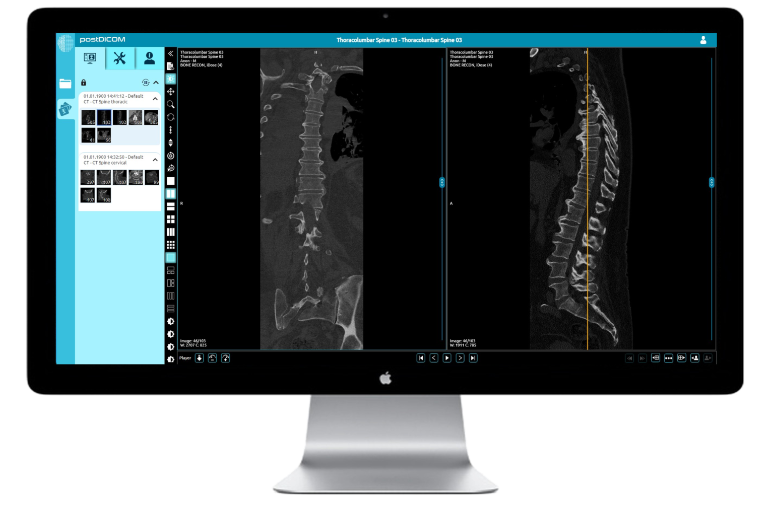
Our imaging courses are very much an interactive experience. Presentations are kept to the minimum and then you'll be into the fully featured cloud based DICOM viewer, looking at cases, feeding back your findings using our interactive tools. You'll get immediate feedback and learning points from our expert faculty member.

DICOM viewer
DICOM viewer
POSTDICOM Logo
Sample the DICOM viewer here. A window will load below the buttons (best demonstrated on a computer rather than mobile device)
Edit Content

Frequently asked questions

We will contact you by email one week before the course takes place with all the necessary links and joining information.

We will re-send the links the day before the course.

If you have not received an email from us please contact us at webinars@infomedltd.co.uk and we will respond ASAP.

NO. Infomed shall provide you, upon registration a link to stream the course within your web browser, or you can download a small application to run it as a separate window on your computer. If you would prefer a mobile device, we shall also include a link download an app from the Play Store/App Store.

YES! It is very much encouraged. There will be Q&A sessions chaired by Infomed. You can type your questions in the 'chat' facility and they will be put to the speakers.

You can find your catch-up in your account page.

At the end of the catch-up page you will find a link to the feedback form, which will generate your CPD certificate when you submit your feedback.

If the catch-up is not visible in your account, please contact us and we will amend your account ASAP.

Using the short videos below, we shall guide you through the process of joining a meeting using Webex. If joining from your own computer If you are connecting from your own device then it is likely that you will be able to join via the Webex application. If joining from a trust/institution computer However, if you are using a computer that is owned and restricted by your trust, then you may find it easier to join via your web browser. Please see the second video for guidance on this process.

Joining Webex using the application on your PC or Mac

Joining Webex using your web browser

Using the short videos below, we shall guide you through the process of opening the PACS and then on to opening, manipulating, and closing a case. You are welcome to access our demo case set below
View demo cases here Password: INFOMED

Accessing the database and cases on PACS​

Advanced features of PACS​

When you connect to a course you should see some introductory slides and hear music.

If you cannot hear any music please check you are connected to the audio.

At the bottom of the webex meeting you may see a button that says "Connect to audio".

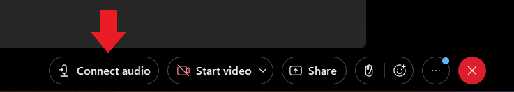
Click this and then select "Use computer for audio" in the pop-up box.

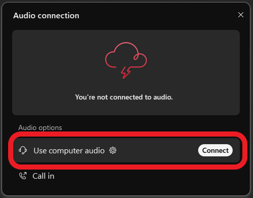
If you have connected by a browser you may need to give your browser access to your microphone in order to connect to the audio.

Click the padlock in the top left of your browser and make sure microphone access is allowed

If this does not resolve your issue please email us or call us on 0204 520 5081

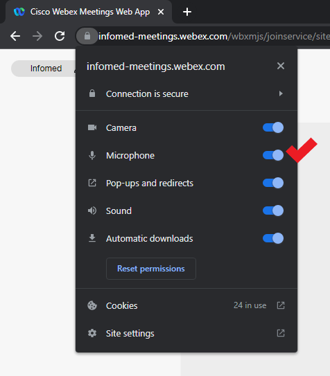
To join an Infomed Online course you simply need an internet connection and a browser (Google Chrome, Mozilla Firefox, Apple Safari).

You can also connect from a mobile device: Download the Webex Meetings app from your App Store.

To join a course with a smooth experience, your internet connection must be stable, not connected to a VPN and at least 20Mbps download.

Below you can use the tool to run an internet speed test.

You must test from:

  • -- the location that you intend the see the course from;
  • -- withing the location, if using Wi-Fi, the room or department area that you intend to view the course from to ensure a good signal
  • -- if connecting from home, a computer that is not connected to a workplace VPN

Internet Speed Test

Please test your connection speed at www.fast.com

To join a course with a smooth experience, your internet connection must be stable, not connected to a VPN and at least 20Mbps download.


Course fees

Live access, plus 90 days access to course recordings and cases.
Three days: £495 | Two days: £325 | One day: £215

Stay up to date with new releases

Subscribe for updates and offers on new events for your specialty.