This event took place recently. You can now:
Abdominal and Pelvic MRI Course
Stream it now with the on-demand catch-up service
- An interactive online course for providing a focused update on abdominal and pelvic MRI. Suitable for all Consultant Radiologists and Senior Trainees
- Format: Short theory lectures followed by interactive interpretation and reporting sessions with your own PACS access.
- CPD: 12 credits with certificate in accordance with the CPD Scheme of the Royal College of Radiologists (RCR), UK

Programme
DAY ONE
60 minutes
Gynaecology – Adnexal lesions and benign disease
Dr Nishat Bharwani
Consultant Radiologist and Hon Clinical Senior Lecturer, Imperial College Healthcare NHS Trust
60 minutes
Gynaecology – Endometrial and cervical cancer
Dr Victoria Stewart
Consultant Radiologist and Hon Clinical Senior Lecturer, Imperial College Healthcare NHS Trust
50 minutes
Renal
Prof Wady Gedroyc
Consultant Radiologist, Imperial College Healthcare NHS Trust, and Chair in Radiology, Imperial College London
60 minutes
Colorectal
Dr Miranda Harvie
Consultant Radiologist, London Northwest Healthcare NHS Trust
70 minutes
Prostate
Dr Amish Lakhani
Consultant Radiologist, Imperial College Healthcare NHS Trust, London and Mount Vernon Hospital
50 minutes
MRA/MRV
Dr Narayan Karunanithy
Consultant Interventional Radiologist and Hon Senior Lecturer, Guy's and St Thomas' NHS Foundation Trust
DAY TWO
60 minutes
Small Bowel
Dr Gurinder Nandra
Consultant Radiologist, Barts Health NHS Trust
60 minutes
Fistula
Dr Catalin Ivan
Consultant Radiologist, Buckinghamshire Healthcare NHS Trust
60 minutes
Liver
Dr Priti Dutta
Consultant Radiologist, Royal Free Hospital
60 minutes
HPB and MRCP
Dr Priti Dutta
Consultant Radiologist, Royal Free Hospital
70 minutes
Edge of the Film: Bone Marrow and Soft Tissue of the Pelvis
Dr Chris Lord
Consultant Radiologist, Imperial College Healthcare NHS Trust
Course director

Dr Nishat Bharwani
Dr Nishat Bharwani is a Consultant Radiologist at Imperial College Healthcare NHS Trust and Honorary Clinical Senior Lecturer at Imperial College London. She qualified with honours from Guy’s, King’s & St Thomas’ Medical School in 2000 and undertook basic medical training in London and Brighton, obtaining membership of the Royal College of Physicians in 2003. She completed general Radiology training at St George's Hospital (2003-2008) and a fellowship in body MRI at Barts and The London NHS Trust (2008-2010), becoming a Fellow of the Royal College of Radiologists in 2008. Her radiological interests include gynaecological imaging, oncological imaging and urological imaging and she has published in these areas. Dr Bharwani is heavily involved in teaching as joint Training Programme Director for Radiology trainees at Imperial, lead for the NW London panel writing MCQs for the FRCR 2A examination, and regularly delivers lectures and workshops both nationally and internationally.
Faculty profiles

Prof Wady Gedroyc
Prof Wady Gedroyc is a Consultant Radiologist at Imperial College Healthcare NHS Trust, Chair in Radiology at Imperial College London, and Medical Director of Magnetic Resonance Imaging at St Mary’s Hospital. He qualified in medicine in 1978 at St Mary's Hospital, London. He undertook radiology training at Guy’s Hospital, London and was an assistant Professor at Penn State University. He has been a full-time consultant at St Mary's since 1990. He has over 90 papers published in peer reviewed journals, mostly on MR related topics. These include multiple research papers on interventional MRI and the development of MRgFUS in various clinical situations. He is an international expert on MR-guided interventional techniques having treated nearly 500 patients with this procedure.

Dr Narayan Karunanithy
Dr Narayan Karunanithy qualified in 1999 with honours from Guy’s, King’s and St Thomas’ Medical School. After completing basic surgical and radiology training, he undertook fellowship training in interventional radiology at Imperial College London and musculoskeletal radiology at the Royal National Orthopaedic Hospital, Stanmore. He was appointed consultant interventional radiologist and honorary clinical lecturer at Guy’s and St Thomas’ in 2010. His specialist interests include: venous diseases; urology, renal, transplant and vascular access radiology in adults and children; and magnetic resonance and computerised tomography angiography. Research interests include applications of non contrast and other novel MR angiographic techniques to image renal/peripheral arteries and deep veins.

Dr Miranda Harvie
Miranda has been a consultant for more than 20 years, having trained in NZ before coming to the UK. The majority of her working life has been based in West London working as a general radiologist with subspecialty interests.
For this course she wants each of you to feel comfortable reviewing colorectal MRI cases, and in particular to have an understanding of rectal cancer MRI staging such that each of you can understand the nuts and bolts of a sub-specialist radiologist’s report, sufficient to be able to review such a case with clinical colleagues as needed e.g. in the absence of the reporter. With luck, a few may be suitably excited by this challenging corner of radiology to use this as a springboard to seek more extensive training, but also to understand the role of MRI in problem solving those occasional colorectal conundrums.
Miranda spends much of her spare time in a 10-pole allotment, which her family have much difficulty removing her from at the weekends.

Dr Amish Lakhani
Dr Amish Lakhani is a Consultant Oncological and Genitourinary Radiologist at Mount Vernon Cancer Centre and Imperial College Healthcare NHS Trust. He is the lead radiologist for the Mount Vernon CyberKnife MDT and a core member of the specialist Uro-oncology MDTs at Imperial.
He completed his medical training at the University of Cambridge and University College London where he graduated with Distinction.
Following completion of his specialist radiology training at Imperial College he was appointed Clinical Fellow in Oncological Imaging at the Royal Marsden NHS Foundation Trust.
He has special interests in whole-body MRI, PET-CT, image-guided radiotherapy planning and multi-parametric prostate MRI.
Dr Lakhani is the radiology training lead at Mount Vernon. He is an invited examiner on several FRCR courses and is Clinical Supervisor for radiology trainees.
He is an Honorary Clinical Senior Lecturer at Imperial College London. He has presented internationally, published in international journals and has been awarded national and international prizes.
The content
- Faculty of practicing consultant radiologists, experts in their fields, with extensive experience in advancing the abdominal and pelvic MRI skills of peers nationally and internationally
- No didactic lectures – practical course, with case based learning and teaching pearls.
- Comprehensive - covering a full range of abdominal and pelvic imaging topics.
- Stimulating, interactive, challenging cases, and very importantly, immediate feedback with around 100 cases to interpret and report
Topics:
- Renal
- Gynaecology – Benign & Malignant
- Colorectal
- Prostate
- MRA/MRV
- Small Bowel
- Fistula
- Liver
- Hepatobiliary/MRCP
The aim
To provide Consultant Radiologists with a practical, stimulating and comprehensive update on advanced interpretation and reporting practice in abdominal pelvic MRI:
- practical (case-based learning);
- stimulating (interactive, challenging cases, immediate feedback)
- comprehensive (covering a full range of abdo and pelvic imaging topics)
By the end of the course, the delegate will have:
- (1) a comprehensive understanding of best practice in advanced abdominal pelvic MRI interpretation and reporting
- (2) improved body MRI interpretation and reporting skills;
- (3) greater confidence in their advanced practice; and
- (4) identified skills and knowledge gaps, if any, relevant to their practice, and clear ways by which these can be addressed.
Comments from 2023
- Thanks, excellent coverage encompassing all important organs and pathologies
- Good number of cases, very interactive, very good lecturers, supportive, friendly
- Very good online platform, quality of streaming, good to have access to recorded teaching.
- I did like the comprehensive contents delivered by expert instructors in interactive learning.
- I thought having Slido was good for interaction and learning.
- This course had a wide variety of cases and pathologies, all clearly presented and good time keeping…and on line.
- Good range of subjects and content and a good PACS and streaming service
- Very good variety of cases and presentations and the ability to interact with the lecturers eg, slido , chat. All good.
- Positive and good update on various topics it met my CPD need
- This course helped me to improve my professional knowledge, and will lead to better diagnosis, reporting and patient care.
- This course has definitely improved diagnostic skills and my confidence for my daily reporting
Comments from 2022
- Coverage within the topics, interactive case review
- Quality of cases
- Gynae sessions, interactive nature, cases
- Broad content, cases, excellent speakers
- the cases, speakers and questions
- good range of topics and pathology. aimed at general radiologist.
- 1) varying levels of depth coverage 2) adequate time to cover the items 3) good coverage of answers
- Lots of cases to view, interaction, smooth pacs
- IT support, easy pacs use, Gyne section
- choice of cases, interaction and overall good lecturers
- Broad coverage, “generalist” approach, use of cases
- PACS, streaming without issues, all lectures good overview
- Online access, quality of lecturers, coverage of topic
Comments from 2021
- Short, good topics, some good lecturers
- Good and relevant level of detail, important/key pathology discussed, good timing and organisation.
- The presenters were all very nice, calm and knowledgeable and were prepared to answer questions or comments in the chat box, so felt very relaxed and personal.
- The support from Infomed was very good all along. Cases
- Comprehensive lectures, perfect teachers and good delivery
- I liked the possibility of having a short presentation/repetition of the important features and the big choice of clinical cases, often picked out with much dexterity
- Very explicative easy to understand better than a usual congress
- I liked the ability to access cases and recording following the course, small group sessions meant participation was easier, broad coverage and working off your own workstation
- I was able to attend the course without travelling.
- I feel much more comfortable to report more complex MRI imaging with a better understanding of the sequences and their uses.
- Great refreshment of knowledge, learned few new things too
- Great course covering lots of important elements of MRI
- Made me more confident in my role as a reporter and to be able to demonstrate abnormalities to surgeons etc for reports outside my reporting area.

Access to cases for our imaging events
Our imaging courses are very much an interactive experience. Presentations are kept to the minimum and then you'll be into the fully featured cloud based DICOM viewer, looking at cases, feeding back your findings using our interactive tools. You'll get immediate feedback and learning points from our expert faculty member.
- Attendance of the course includes access to the database of cases associated to this event on our server at PostDICOM.
- Full access to each case with a full toolset to open, view and manipulate each case alongside the faculty but on your own screen!
- You will maintain your access to the resource throughout your 60 day catch-service period too.
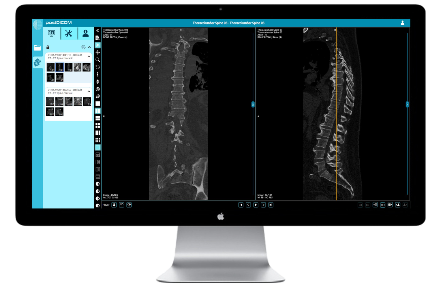
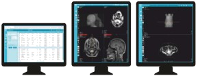

Sample the DICOM viewer here. A window will load below the buttons (best demonstrated on a computer rather than mobile device)
We will contact you by email one week before the course takes place with all the necessary links and joining information.
We will re-send the links the day before the course.
If you have not received an email from us please contact us at webinars@infomedltd.co.uk and we will respond ASAP.
NO. Infomed shall provide you, upon registration a link to stream the course within your web browser, or you can download a small application to run it as a separate window on your computer. If you would prefer a mobile device, we shall also include a link download an app from the Play Store/App Store.
YES! It is very much encouraged. There will be Q&A sessions chaired by Infomed. You can type your questions in the 'chat' facility and they will be put to the speakers.
You can find your catch-up in your account page.
At the end of the catch-up page you will find a link to the feedback form, which will generate your CPD certificate when you submit your feedback.
If the catch-up is not visible in your account, please contact us and we will amend your account ASAP.
Joining Webex using the application on your PC or Mac
Joining Webex using your web browser
View demo cases here Password: INFOMED
Accessing the database and cases on PACS
Advanced features of PACS
When you connect to a course you should see some introductory slides and hear music.
If you cannot hear any music please check you are connected to the audio.
At the bottom of the webex meeting you may see a button that says "Connect to audio".
Click this and then select "Use computer for audio" in the pop-up box.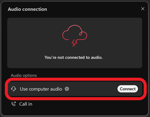
If you have connected by a browser you may need to give your browser access to your microphone in order to connect to the audio.
Click the padlock in the top left of your browser and make sure microphone access is allowed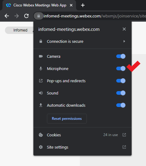
If this does not resolve your issue please email us or call us on 0204 520 5081
To join an Infomed Online course you simply need an internet connection and a browser (Google Chrome, Mozilla Firefox, Apple Safari).
You can also connect from a mobile device: Download the Webex Meetings app from your App Store.
To join a course with a smooth experience, your internet connection must be stable, not connected to a VPN and at least 20Mbps download.
Below you can use the tool to run an internet speed test.
You must test from:
- -- the location that you intend the see the course from;
- -- withing the location, if using Wi-Fi, the room or department area that you intend to view the course from to ensure a good signal
- -- if connecting from home, a computer that is not connected to a workplace VPN
Internet Speed Test
Please test your connection speed at www.fast.com
To join a course with a smooth experience, your internet connection must be stable, not connected to a VPN and at least 20Mbps download.
Course fees
£295 (inc. VAT)
- 120 days of access with unlimited playback during this time
- CPD Certificate of attendance upon completion with 12 CPD points
- Full access and control to the DICOM cases
- Lots of cases to review and interpret
