For all General Consultant Radiologists and Senior ST Radiologists
Sessions are pitched at a level suitable for the Consultant Radiologist
Established, 2004 in the UK
CPD accredited, online courses for doctors in all of the major specialties
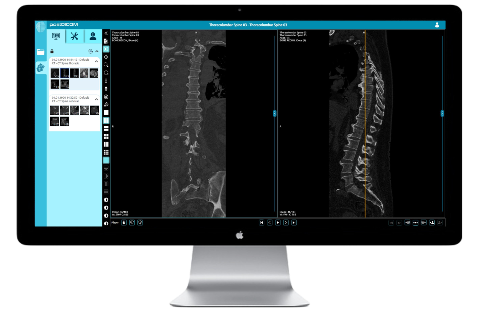
Attendees will be provided with individual, secure access to the cases and advanced image viewing PACS software so that they can go through the cases with the faculty, simultanteously.
Attendees will be asked to evaluate each session and will be provided with a CPD certificate upon completion This course provides 12 CPD credits in accordance with the CPD Scheme of the Royal College of Radiologists
Purchasing the catch-up will give you access to the webinar recordings for 90 days. You'll also maintain your access to the cases during this time too
Our imaging courses are very much an interactive experience. Presentations are kept to the minimum and then you'll be into the fully featured cloud based DICOM viewer, looking at cases, feeding back your findings using our interactive tools. You'll get immediate feedback and learning points from our expert faculty member.
For all General Consultant Radiologists and Senior ST Radiologists
Sessions are pitched at a level suitable for the Consultant Radiologist
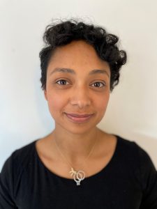
Consultant Radiologist, Royal Free Hospital, London
Dr Dutta is a consultant interventional radiologist at the Royal Free Hospital in London. She previously served as a consultant interventional radiologist at the Lister Hospital in Stevenage, Hertfordshire.
Dr Dutta studied medicine at University College London (UCL), graduating in 2008 with an intercalated BSc in neuroscience.
Her current interests are centred on liver imaging and intervention. She plays an active role in the hospital's regional multidisciplinary team meetings which encompass a wide case mix of hepatobiliary, pancreatic and neuroendocrine tumours, as well as colorectal metastases. She has published on topics including arteriovenous fistula intervention and the relationship of sarcopenia to outcomes in EVAR patients.
She is currently publishing work related to the follow-up of indeterminate liver lesions in cirrhotic patients.
Her day-to-day practice with relation to liver intervention ranges from, transjugular liver biopsies, transarterial liver embolization, liver ablation, portal vein embolization, palliative stenting, TIPS placement and embolization of bleeding varices.
Twelve sessions over two days:
Each session includes:
To provide Radiologists with a practical, stimulating and comprehensive update on advanced interpretation and reporting practice in HPB:
By the end of the course, the delegate will have:
70 mins
50 mins
80 mins
50 mins
50 mins
50 mins
60 mins
60 mins
60 mins
60 mins
50 mins
50 mins
40 mins
NO. Infomed shall provide you, upon registration a link to stream the course within your web browser, or you can download a small application to run it as a separate window on your computer. If you would prefer a mobile device, we shall also include a link download an app from the Play Store/App Store.
YES! It is very much encouraged. There will be Q&A sessions chaired by Infomed. You can type your questions in the ‘chat’ facility and they will be put to the speakers.
YES! We shall ask for your evaluation of the faculty and organisation. Upon completing this, we shall send you a certificate of attendance by email.

© 2023 All rights reserved